
Non-Compression Cone Beam Breast CT
What Is Cone Beam Breast CT?
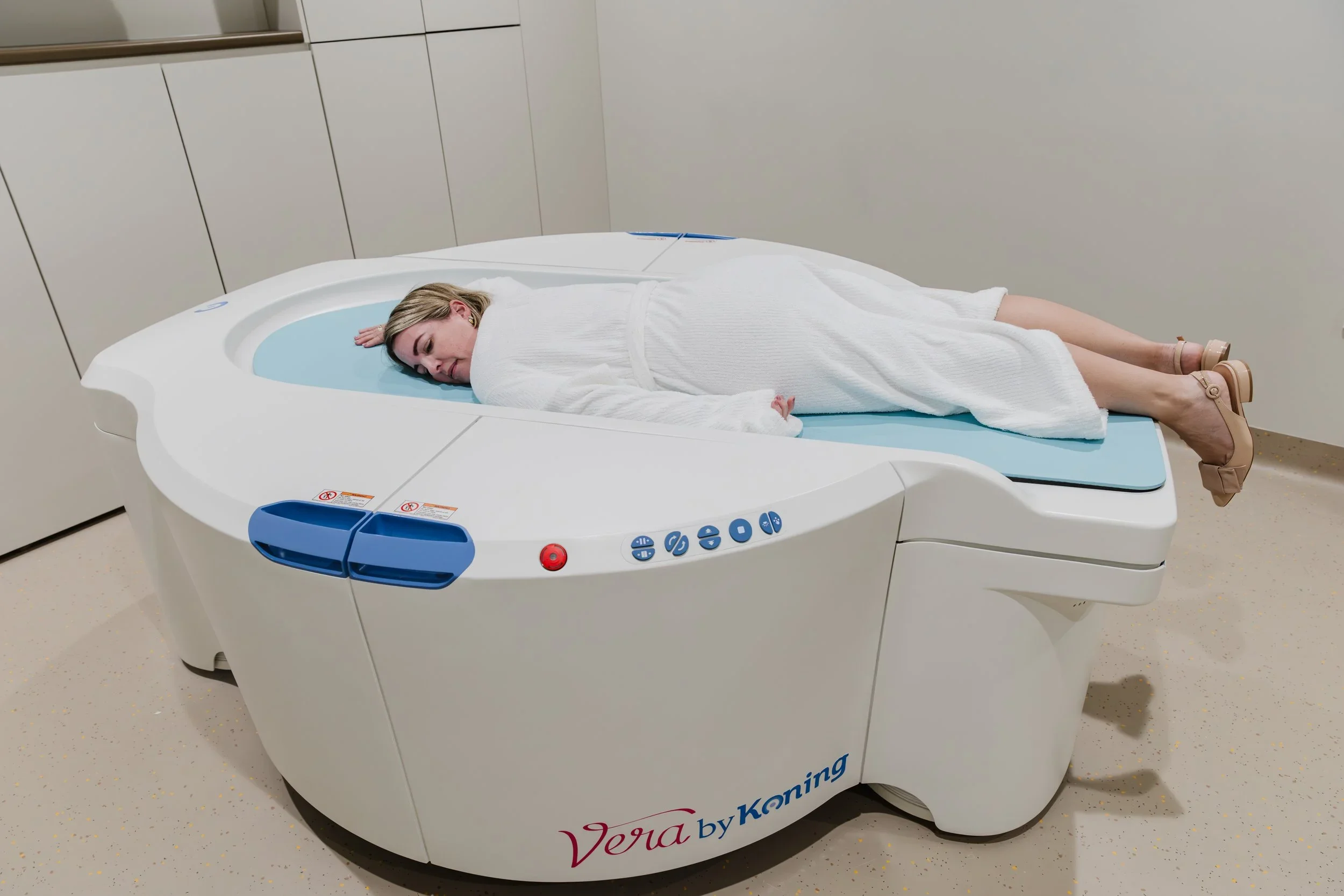
Cone Beam Computed Tomography (CBCT) is an advanced imaging technique used to take detailed 3D images of the breast.
Unlike traditional mammography, which provides 2D images, CBCT offers a comprehensive view of breast tissue in three dimensions, helping doctors detect and diagnose abnormalities more effectively.
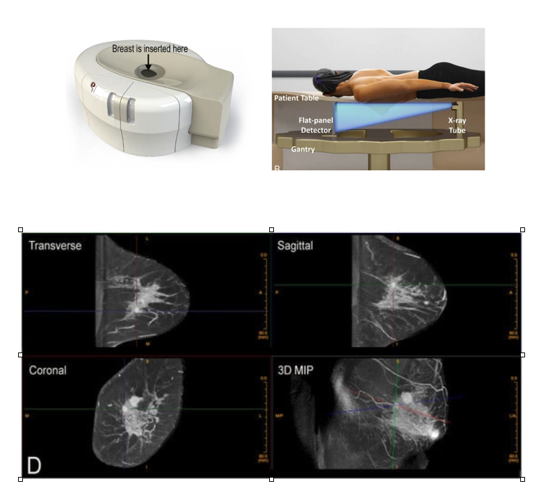
Cone-beam breast CT can provide detailed 3D imaging without breast compression and has applications in the assessment of breast lesions, biopsy guidance and surgical planning.
How Does CBCT Work?
During a CBCT scan:
Positioning
You will lie face down on a specially designed table with an opening for the breast being examined.
No Compression
The breast is positioned in the imaging area, with no compression needed, making the procedure more comfortable than a mammogram.
Imaging
The scanner rotates around the breast, capturing detailed 3D images using low-dose X-rays.
Scan Time
The entire procedure typically takes about 10 minutes.
Why Might You Need a CBCT?
CBCT is particularly useful for:
Women with dense breast tissue, where abnormalities may be harder to detect on mammograms.
Further evaluation of an abnormal finding on a mammogram or ultrasound.
Providing highly detailed images to guide biopsies or surgical planning.
Cone-beam breast CT can also provide information about breast density and tumour characteristics.
Benefits of CBCT for Breast Imaging
Comfort
No breast compression is required.
Accuracy
Provides a 3D view, reducing overlap of breast tissue that can obscure abnormalities on standard mammograms.
Efficiency
Quickly captures high-resolution images.
Low Radiation Dose
Uses a lower dose of radiation compared to traditional CT scans.
Are There Any Risks?
Radiation Exposure
CBCT uses X-rays, but the radiation dose is low and considered safe.
Not a Screening Tool
CBCT is not typically used as a routine screening method but as a supplementary tool for specific cases.
Frequently Asked Questions
-
Cone Beam Computed Tomography (CBCT) is an advanced imaging technique used to take detailed 3D images of the breast.
Unlike traditional mammography, which provides 2D images, CBCT offers a comprehensive view of breast tissue in three dimensions.
-
No.
The breast is positioned in the imaging area without compression, making the procedure more comfortable than a mammogram.
-
You will lie face down on a specially designed table with an opening for the breast being examined.
The scanner rotates around the breast, capturing detailed 3D images using low-dose X-rays.
-
The entire procedure typically takes about 10 minutes.
-
CBCT may be useful for women with dense breast tissue, further evaluation of an abnormal finding on a mammogram or ultrasound, or to provide highly detailed images to guide biopsies or surgical planning.
-
Benefits include no breast compression, 3D imaging that reduces the overlap of breast tissue, quick acquisition of high-resolution images and a low radiation dose compared to traditional CT scans.
-
CBCT uses X-rays, but the radiation dose is low and considered safe.
-
CBCT is not typically used as a routine screening method but as a supplementary tool for specific cases.
Helpful Resources
Need Further Advice?
If you have questions about whether Cone Beam CT is appropriate for you, speak with your healthcare provider or breast imaging specialist.
Your healthcare team can help determine which imaging method is best suited to your individual circumstances.