
Contrast Enhanced Mammography
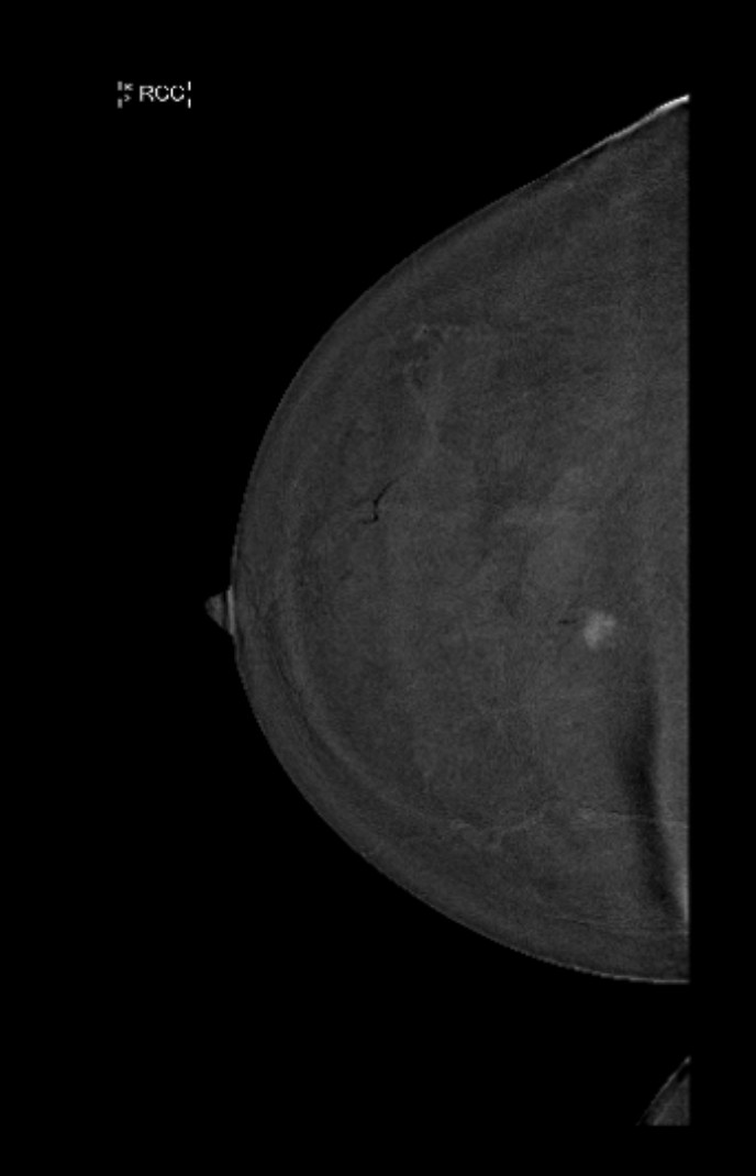
What Is Contrast Enhanced Mammography?
Contrast Enhanced Mammography is a new and exciting addition to traditional 3D mammography that can improve the detection of small early breast cancer and help identify breast cancers in dense breast tissue that may not be visible on routine conventional mammography.
Contrast Enhanced Mammography uses iodine-based contrast to identify cancers that are typically vascular and have an increased blood flow.
Computer algorithms can distinguish between cancerous tissue that absorbs the iodine contrast and dense breast tissue that absorbs the contrast to a lesser extent.
The computer systems allow the non-cancerous breast tissue to be subtracted from the image, making the concerning cancerous tissue more obvious.
This technique is called Dual Energy Subtraction (DES).
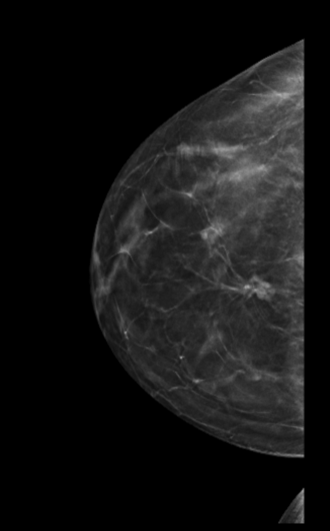
Standard Conventional Breast Imaging - Previous Breast Surgery with Scarring
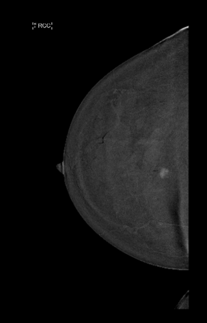
Contrast Enhanced Mammogram with Dual Energy Subtraction - Scar tissue is removed and the cancer is clearly identified as a white spot
What Are the Benefits of Contrast Enhanced Mammography?
Contrast Enhanced Mammography is beneficial in dense breast tissue and in breasts that have had prior surgery.
Dense breast tissue and previous scar tissue typically obscure small cancers.
Cancer and scar tissue can be very difficult to distinguish and usually requires a biopsy.
Contrast Enhanced Mammography can help distinguish between benign scar tissue, dense breast tissue and cancers.
This allows only those patients who really need a biopsy to undergo a needle biopsy.
Contrast Enhanced Mammography and Dense Breast Tissue
MRI is often recommended in dense breast tissue as the dense tissue makes identification of small cancers difficult.
MRI are challenging as they need to be timed to patients menstrual cycle, takes 30-40 minutes and can be quite claustrophobic.
Contrast enhanced mammography can identify tumour with additional accuracy and only requires an additional 10-15mins of time.
How Is Contrast Mammography Performed?
Contrast Enhanced Mammography requires an IV drip to be placed.
A standard conventional mammogram is initially performed.
This will be followed by the contrast enhanced images.
A small dose of iodine-based contrast will be injected immediately prior to the enhanced images being taken.
This allows time for the contrast to circulate to the breast prior to the images being taken.
When Is Contrast Mammography Used?
Contrast mammography is now routinely offered on all of Dr Greens post breast cancer surveillance patients, in the screening setting where the breasts are dense or complex and in high-risk patients undergoing regular surveillance screening.
Need Further Advice?
If you have questions about whether Contrast Enhanced Mammography, speak with your healthcare provider or breast imaging specialist.