
B3 Breast Lesions and Excisional Biopsy

Understanding B3 Breast Lesions
B3 breast lesions are a group of abnormal but non-cancerous (benign) breast tissue changes detected during a breast biopsy. The term “B3” refers to the classification used to categorise these lesions, which have uncertain or borderline malignant potential.
While B3 lesions are not cancer, they may carry a higher risk of developing into breast cancer or being associated with cancer in nearby tissue.
What are B3 Breast Lesions?
B3 breast lesions include several different types of abnormal tissue changes. These findings are often detected during routine mammograms or other breast imaging tests and confirmed by biopsy.
B3 breast lesions include:
Atypical Ductal Hyperplasia (ADH)
Abnormal growth of cells within the milk ducts.
Lobular Neoplasia (LN)
Abnormal cells in the lobules, also called atypical lobular hyperplasia or lobular carcinoma in situ (LCIS).
Papillomas
Small, wart-like growths inside the ducts, sometimes containing abnormal cells.
Radial Scars (Complex Sclerosing Lesions)
Star-shaped areas of scar-like tissue.
Flat Epithelial Atypia (FEA)
Abnormal cells lining the ducts.
Who is at Risk?
The exact cause of B3 lesions is unknown, and anyone can develop them. However, certain risk factors may increase the likelihood.
These include:
Age: B3 lesions are more common in women over 40.
Hormonal factors: Some hormonal changes may play a role in the development of B3 lesions.
Family history: Women with a family history of breast cancer may have a higher risk.
How are B3 Lesions Diagnosed?
B3 lesions are usually detected during imaging tests, often before any symptoms appear.
The diagnosis process includes:
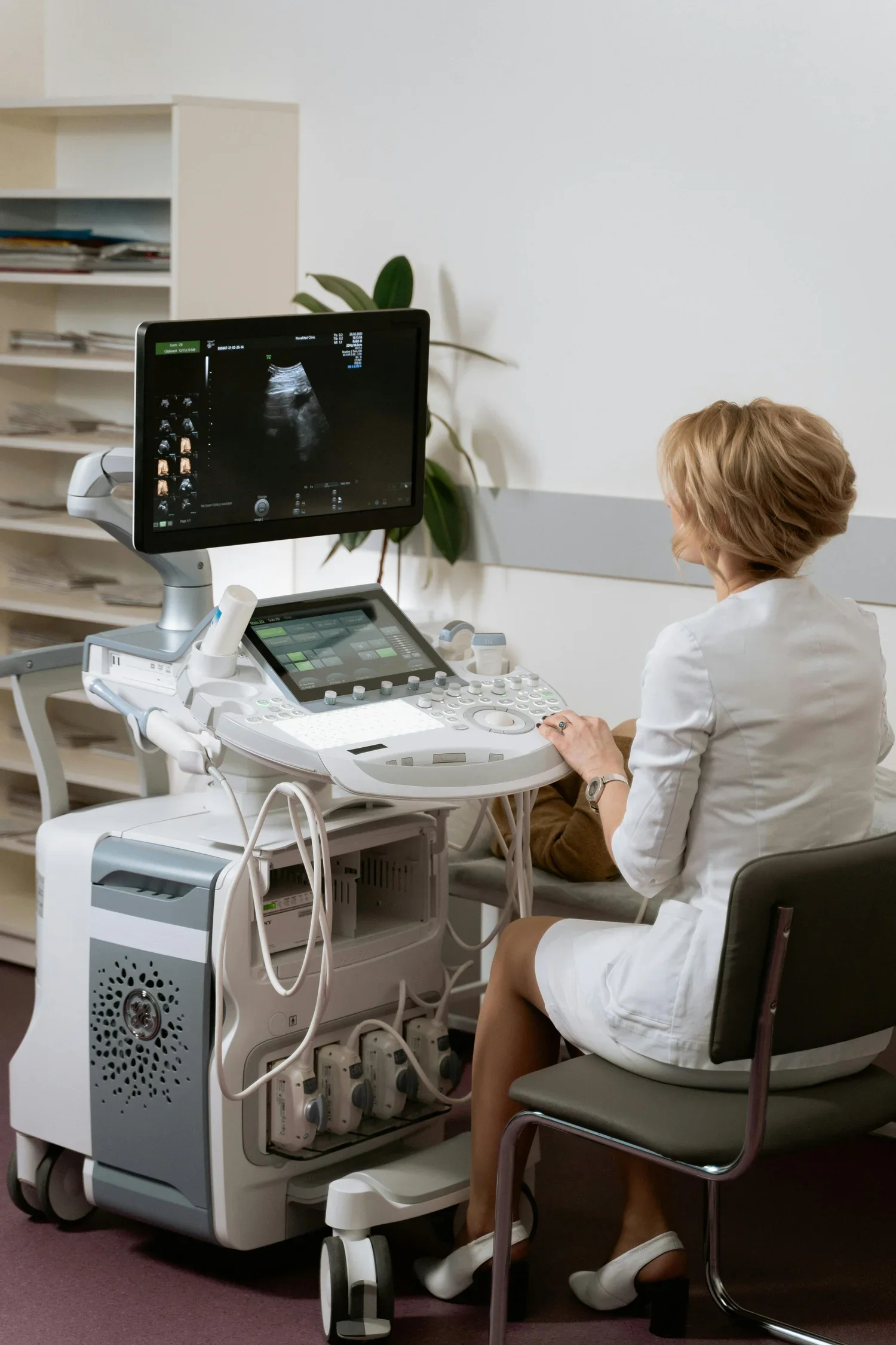
Mammogram or Ultrasound
B3 lesions may appear as suspicious areas on a mammogram or ultrasound.
Core Needle Biopsy
A tissue sample is taken from the suspicious area to confirm whether the lesion is B3.
Pathology Analysis
A pathologist examines the biopsy sample to determine the exact type of lesion and assess the presence of any abnormal or atypical cells.
What are the Treatment Options for B3 Lesions?
Treatment for B3 lesions varies depending on the lesion type, size, and the presence of any atypical or abnormal cells.
Options include surgical excision, close monitoring (surveillance) and hormone therapy.
Surgical Excision
Surgical removal (excision) of the lesion may be recommended to ensure all atypical cells are removed and to check for any signs of early cancer in the surrounding tissue.
This is often the case for lesions with higher risk features, such as atypical ductal hyperplasia or lobular neoplasia.
Close Monitoring (Surveillance)
In cases where the B3 lesion has low-risk features, close monitoring may be an option.
This includes regular imaging tests, such as mammograms or MRIs, and follow-up clinical exams. Monitoring is usually done every 6 to 12 months.
Hormone Therapy
For some patients, especially those with lobular neoplasia, hormone therapy, such as tamoxifen, may be considered to help reduce the risk of developing breast cancer.
This option is often discussed if there are additional risk factors
Treatment for B3 lesions varies depending on the lesion type, size, and the presence of any atypical or abnormal cells.
Options include surgical excision, close monitoring (surveillance) and hormone therapy.

The Excisional Breast Biopsy Procedure
Consultation
During your consultation, your surgeon will review your breast imaging and biopsy results and discuss the type of B3 lesion that has been identified.
Your goals, concerns and treatment options will be discussed, including whether surgical excision is recommended based on the type of lesion, its features and your individual circumstances.
Surgery
An excisional breast biopsy involves the surgical removal of the abnormal area of breast tissue.
Surgical removal may be recommended to ensure all atypical cells are removed and to check for any signs of early cancer in the surrounding tissue.
The removed tissue is sent to a pathologist for examination to determine the exact nature of the lesion and assess the presence of any abnormal or atypical cells.
Recovery
Your surgeon will provide specific instructions regarding your recovery following an excisional breast biopsy.
The recovery period will depend on the extent and location of the procedure and your individual circumstances.
Benefits of Excisional Biopsy
Surgical excision of a B3 lesion may:
Remove the abnormal breast tissue.
Ensure all atypical cells are removed.
Allow the removed tissue to be examined more extensively by a pathologist.
Check for any signs of early cancer in the surrounding tissue.
Your surgeon will discuss whether excision is appropriate for you based on the type of B3 lesion, its features and your individual circumstances.
Risks & Complications
As with any surgical procedure, an excisional breast biopsy carries some risks.
Your surgeon will discuss the potential risks and complications with you during your consultation, including those associated with surgery, wound healing, infection, bleeding, scarring and changes to the breast.
Follow-Up Care
Follow-up care is important for managing B3 lesions.
Your healthcare provider will develop a personalised follow-up plan that may include:
Regular imaging tests: Routine mammograms or other imaging to monitor for changes.
Physical exams: Clinical breast exams to check for any new lumps or changes.
Discussion of lifestyle factors: Maintaining a healthy lifestyle, including regular exercise, a balanced diet and limiting alcohol, may help reduce risk.
Follow-up care will depend on the type of B3 lesion, your individual risk factors and whether the lesion has been surgically removed.
Living with a B3 Lesion Diagnosis
A B3 lesion diagnosis can be concerning, but it is important to remember that these lesions are not cancer.
However, staying proactive with follow-up care is essential to detect any changes early. Many people with B3 lesions never develop breast cancer, but regular monitoring is essential.
Staying informed and vigilant with follow-up care can help ensure the best possible outcomes. Your healthcare team is here to support you with all the information you need to make the best decisions for your health.
Frequenty Asked Questions
-
No. B3 lesions are not cancer. They are abnormal but non-cancerous (benign) breast tissue changes that have uncertain or borderline malignant potential.
However, some B3 lesions may carry a higher risk of developing into breast cancer or being associated with cancer in nearby tissue.
-
This depends on the type of B3 lesion, its size and the presence of any atypical or abnormal cells.
Surgical excision may be recommended for lesions with higher-risk features, while close monitoring may be appropriate for some lesions with low-risk features.
-
B3 lesions are often detected during routine mammograms or other breast imaging tests, sometimes before any symptoms appear.
A core needle biopsy is then used to obtain a tissue sample for pathology analysis
-
Follow-up care is important for managing B3 lesions. Your healthcare provider will develop a personalised follow-up plan that may include regular mammograms, MRI or other imaging tests, as well as clinical breast examinations.
-
B3 lesions are not cancer, but some may carry a higher risk of developing into breast cancer or being associated with cancer in nearby tissue.
The level of risk varies depending on the specific type of B3 lesion and individual circumstances.
-
Maintaining a healthy lifestyle, including regular exercise, a balanced diet and limiting alcohol, may help reduce the risk of developing breast cancer.
-
You may wish to ask:
What type of B3 lesion do I have, and what does it mean?
What follow-up care or treatment do you recommend for me?
Are there lifestyle changes I should consider to reduce my risk?
Is there anything specific I should monitor for at home and after treatment?
How often will I need follow-up exams?
What lifestyle changes should I consider during and after treatment?
Helpful Resources
Need Further Advice?
If you have been diagnosed with a B3 breast lesion, the team at Brisbane Breast and Surgical Care can provide a comprehensive assessment of your biopsy and imaging results and discuss the most appropriate management options for you.
Your healthcare team is here to support you with all the information you need to make informed decisions about your breast health.